Pyelonephritis: Kidney Infection
Contents
Who is at risk for pyelonephritis?
What are the symptoms of pyelonephritis?
What are the complications of pyelonephritis?
How is pyelonephritis diagnosed?
How is pyelonephritis treated?
What is pyelonephritis?
Pyelonephritis is a type of urinary tract infection (UTI) that affects one or both kidneys.
What is the urinary tract?
The urinary tract is the body’s drainage system for removing wastes and extra water. The urinary tract includes two kidneys, two ureters, a bladder, and a urethra. The kidneys are two bean-shaped organs, each about the size of a fist. They are located near the middle of the back, just below the rib cage, one on each side of the spine. Every day, the two kidneys process about 200 quarts of blood to produce about 1 to 2 quarts of urine, composed of wastes and extra water. Children produce less urine than adults. The amount produced depends on their age. The urine flows from the kidneys to the bladder through tubes called the ureters. The bladder stores urine until releasing it through urination. When the bladder empties, urine flows out of the body through a tube called the urethra at the bottom of the bladder.
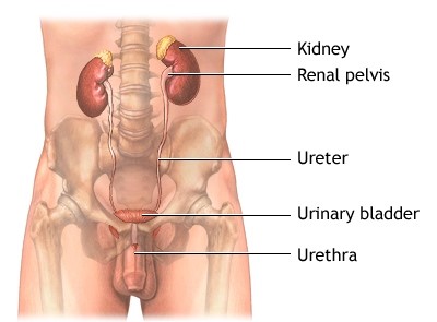
Male urinary system
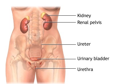
Female urinary system
What causes pyelonephritis?
Pyelonephritis is caused by a bacterium or virus infecting the kidneys. Though many bacteria and viruses can cause pyelonephritis, the bacterium Escherichia coli is often the cause. Bacteria and viruses can move to the kidneys from the bladder or can be carried through the bloodstream from other parts of the body. A UTI in the bladder that does not move to the kidneys is called cystitis.
Who is at risk for pyelonephritis?
People most at risk for pyelonephritis are those who have a bladder infection and those with a structural, or anatomic, problem in the urinary tract. Urine normally flows only in one direction—from the kidneys to the bladder. However, the flow of urine may be blocked in people with a structural defect of the urinary tract, a kidney stone, or an enlarged prostate—the walnut-shaped gland in men that surrounds the urethra at the neck of the bladder and supplies fluid that goes into semen. Urine can also back up, or reflux, into one or both kidneys. This problem, which is called vesicoureteral reflux (VUR), happens when the valve mechanism that normally prevents backward flow of urine is not working properly. VUR is most commonly diagnosed during childhood. Pregnant women and people with diabetes or a weakened immune system are also at increased risk of pyelonephritis.
What are the symptoms of pyelonephritis?
Symptoms of pyelonephritis can vary depending on a person’s age and may include the following:
Children younger than 2 years old may only have a high fever without symptoms related to the urinary tract. Older people may not have any symptoms related to the urinary tract either; instead, they may exhibit confusion, disordered speech, or hallucinations.
What are the complications of pyelonephritis?
Most people with pyelonephritis do not have complications if appropriately treated with bacteria-fighting medications called antibiotics.
In rare cases, pyelonephritis may cause permanent kidney scars, which can lead to chronic kidney disease, high blood pressure, and kidney failure. These problems usually occur in people with a structural problem in the urinary tract, kidney disease from other causes, or repeated episodes of pyelonephritis.
Infection in the kidneys may spread to the bloodstream—a serious condition called sepsis—though this is also uncommon.
How is pyelonephritis diagnosed?
The tests used to diagnose pyelonephritis depend on the patient’s age, gender, and response to treatment and include the following:
- Urinalysis. Urinalysis is testing of a urine sample. The urine sample is collected in a special container in a health care provider’s office or commercial facility and can be tested in the same location or sent to a lab for analysis. The presence of white blood cells and bacteria in the urine indicate infection.
- Urine culture. A urine culture is performed by placing part of a urine sample in a tube or dish with a substance that encourages any bacteria present to grow. The urine sample is collected in a special container in a health care provider’s office or commercial facility and sent to a lab for culture. Once the bacteria have multiplied, which usually takes 1 to 3 days, they can be identified. The health care provider can then determine the best treatment.
- Ultrasound. Ultrasound uses a device, called a transducer, that bounces safe, painless sound waves off organs to create an image of their structure. The procedure is performed in a health care provider’s office, outpatient center, or hospital by a specially trained technician, and the images are interpreted by a radiologist—a doctor who specializes in medical imaging; anesthesia is not needed. The images can show obstructions in the urinary tract. Ultrasound is often used for people who do not respond to treatment within 72 hours.
- Computerized tomography (CT) scan. CT scans use a combination of x rays and computer technology to create three-dimensional (3-D) images. A CT scan may include the injection of a special dye, called contrast medium. CT scans require the person to lie on a table that slides into a tunnel-shaped device where the x rays are taken. The procedure is performed in an outpatient center or hospital by an x-ray technician, and the images are interpreted by a radiologist. Anesthesia is not needed. CT scans can show obstructions in the urinary tract. The test is often used for people who do not respond to treatment within 72 hours.
- Voiding cystourethrogram (VCUG). A VCUG is an x-ray image of the bladder and urethra taken while the bladder is full and during urination, also called voiding. The procedure is performed in an outpatient center or hospital by an x-ray technician supervised by a radiologist, who then interprets the images. Anesthesia is not needed, but sedation may be used for some people. The bladder and urethra are filled with contrast medium to make the structures clearly visible on the x-ray images. The x-ray machine captures images of the contrast medium while the bladder is full and when the person urinates. This test can show abnormalities of the inside of the urethra and bladder and is usually used to detect VUR in children.
- Digital rectal examination (DRE). A DRE is a physical exam of the prostate that is performed in the health care provider’s office. Anesthesia is not needed. To perform the exam, the health care provider asks the person to bend over a table or lie on his side while holding his knees close to his chest. The health care provider slides a gloved, lubricated finger into the rectum and feels the part of the prostate that lies in front of the rectum. Men with suspected pyelonephritis may have a DRE to determine whether a swollen prostate may be obstructing the neck of the bladder.
- Dimercaptosuccinic acid (DMSA) scintigraphy. DMSA scintigraphy is an imaging technique that relies on the detection of small amounts of radiation after injection of radioactive material. Because the dose of radioactive material is small, the risk of causing damage to cells is low. The procedure is performed in an outpatient center or hospital by a specially trained technician, and the images are interpreted by a radiologist. Anesthesia is not needed. Radioactive material is injected into a vein in the person’s arm and travels through the body to the kidneys. Special cameras and computers are used to create images of the radioactive material as it passes through the kidneys. The radioactive material makes the parts of the kidney that are infected or scarred stand out on the image. DMSA scintigraphy is used to show the severity of kidney infection or kidney damage, such as scarring.
How is pyelonephritis treated?
Pyelonephritis is treated with antibiotics, which may need to be taken for several weeks. While a urine sample is sent to a lab for culture, the health care provider may begin treatment with an antibiotic that fights the most common types of bacteria. Once culture results are known and the bacteria is clearly identified, the health care provider may switch the antibiotic to one that more effectively targets the bacteria. Antibiotics may be given through a vein, orally, or both. Urinary tract obstructions are often treated with surgery.
Severely ill patients may be hospitalized and limited to bed rest until they can take the fluids and medications they need on their own. Fluids and medications may be given intravenously during this time.
In adults, repeat urine cultures should be performed after treatment has ended to make sure the infection does not recur. If a repeat test shows infection, another 14-day course of antibiotics is prescribed; if infection recurs again, antibiotics are prescribed for 6 weeks.
Eating, diet, and nutrition have not been shown to play a role in causing or preventing pyelonephritis.
- Pyelonephritis is a type of urinary tract infection that affects one or both kidneys.
- Pyelonephritis is caused by a bacterium or virus infecting the kidneys. Though many bacteria and viruses can cause pyelonephritis, the bacterium Escherichia coli is often the cause. Bacteria and viruses can move to the kidneys from the bladder or can be carried through the bloodstream from other parts of the body.
- Symptoms of pyelonephritis can vary depending on a person’s age and may include the following:
- Children younger than 2 years old may only have a high fever without symptoms related to the urinary tract. Older people may not have any symptoms related to the urinary tract either; instead, they may exhibit confusion, disordered speech, or hallucinations.
- Most people with pyelonephritis do not have complications if appropriately treated with bacteria-fighting medications called antibiotics.
The National Institute of Diabetes and Digestive and Kidney Diseases (NIDDK) and other components of the National Institutes of Health (NIH) conduct and support research into many diseases and conditions.
What are clinical trials, and are they right for you?
Clinical trials are part of clinical research and at the heart of all medical advances. Clinical trials look at new ways to prevent, detect, or treat disease. Researchers also use clinical trials to look at other aspects of care, such as improving the quality of life for people with chronic illnesses.
Source: http://kidney.niddk.nih.gov/kudiseases/pubs/pyelonephritis/